
Cell Diagram Labeled Simple SexiezPicz Web Porn
Unit 1 Intro to biology Unit 2 Chemistry of life Unit 3 Water, acids, and bases Unit 4 Properties of carbon Unit 5 Macromolecules Unit 6 Elements of life Unit 7 Energy and enzymes Unit 8 Structure of a cell Unit 9 More about cells Unit 10 Membranes and transport Unit 11 More about membranes Unit 12 Cellular respiration Unit 13 Photosynthesis

Label the Parts of the Plant and Animal Cell Biology LibreTexts
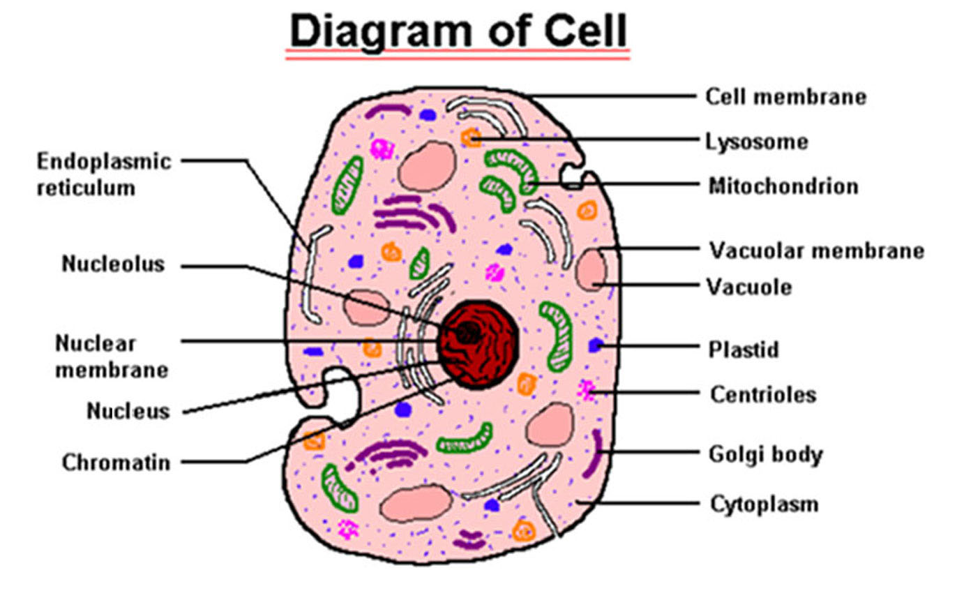
A Labeled Diagram of the Animal Cell and its Organelles There are two types of cells - Prokaryotic and Eucaryotic. Eukaryotic cells are larger, more complex, and have evolved more recently than prokaryotes. Where, prokaryotes are just bacteria and archaea, eukaryotes are literally everything else.

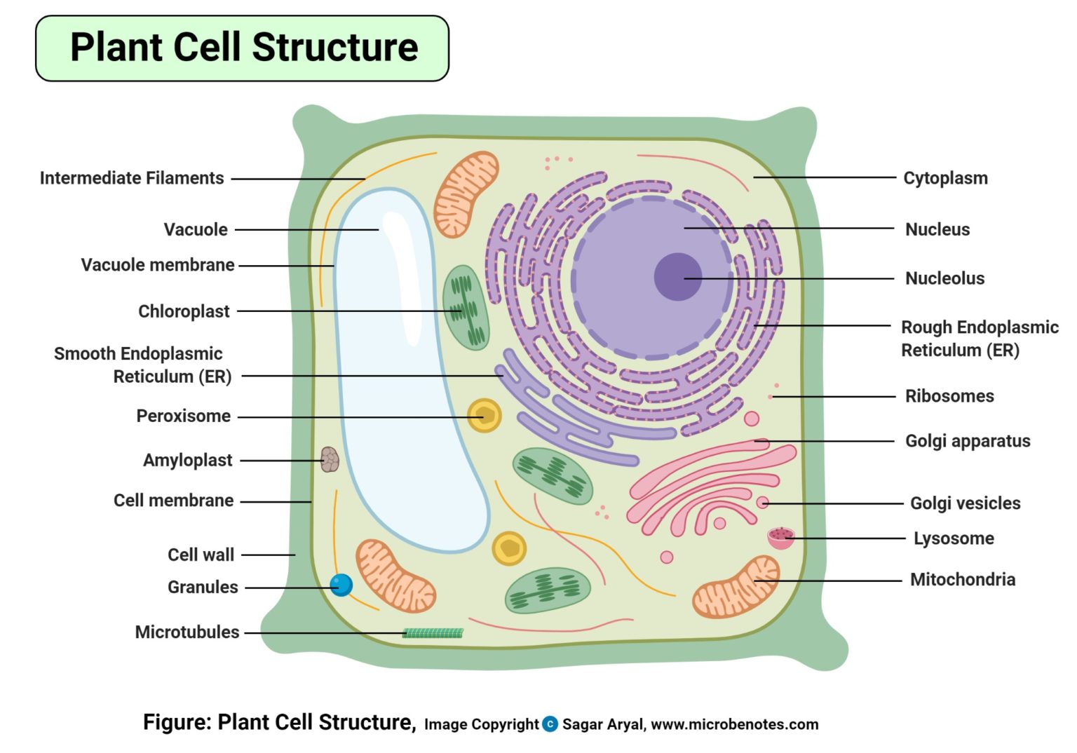
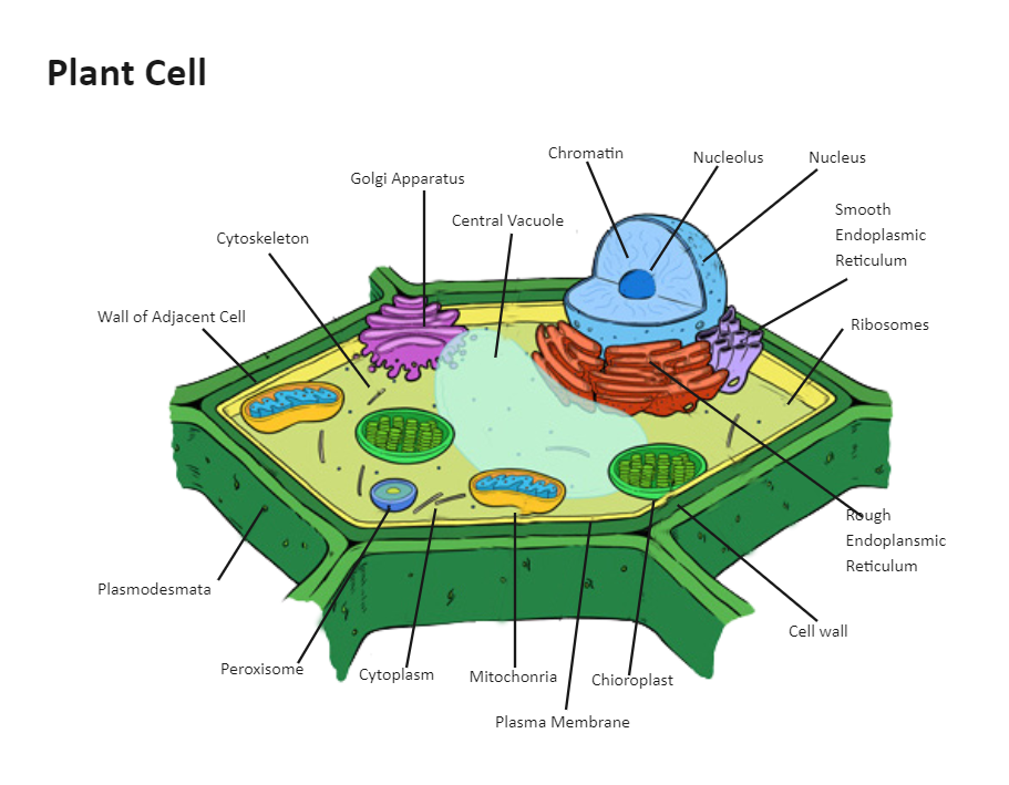
A Labeled Diagram of the Plant Cell and Functions of its Organelles
Cell organelles are specialized entities present inside a particular type of cell that performs a specific function. There are various cell organelles, out of which, some are common in most types of cells like cell membranes, nucleus, and cytoplasm. However, some organelles are specific to one particular type of cell-like plastids and cell.
Printable Animal Cell Diagram Labeled, Unlabeled, and Blank
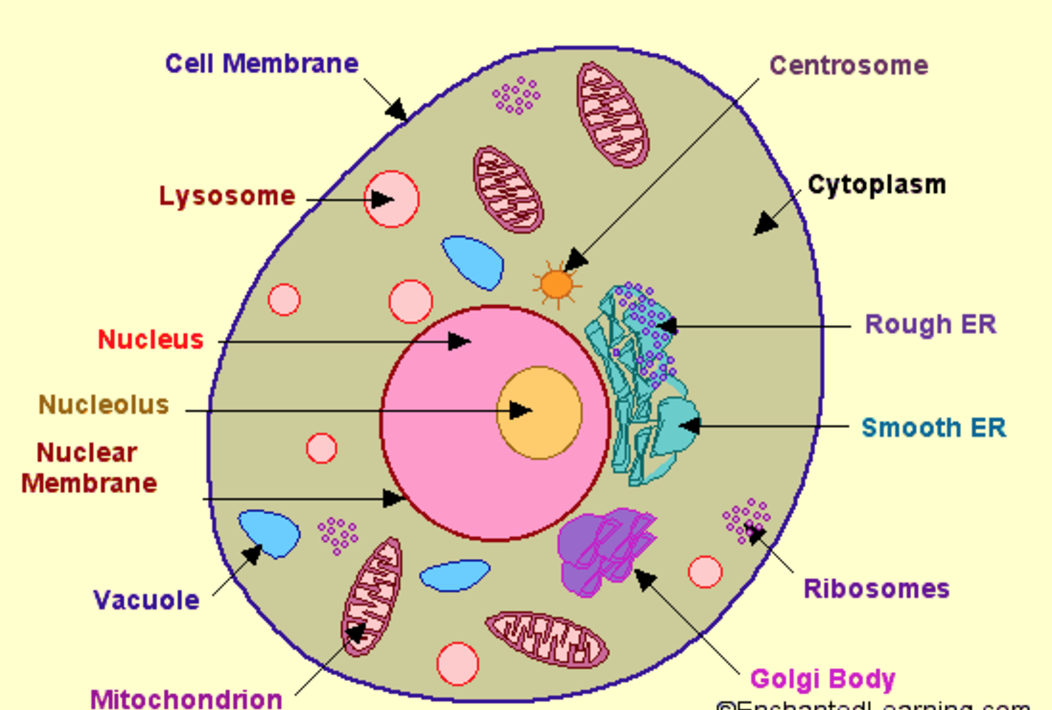
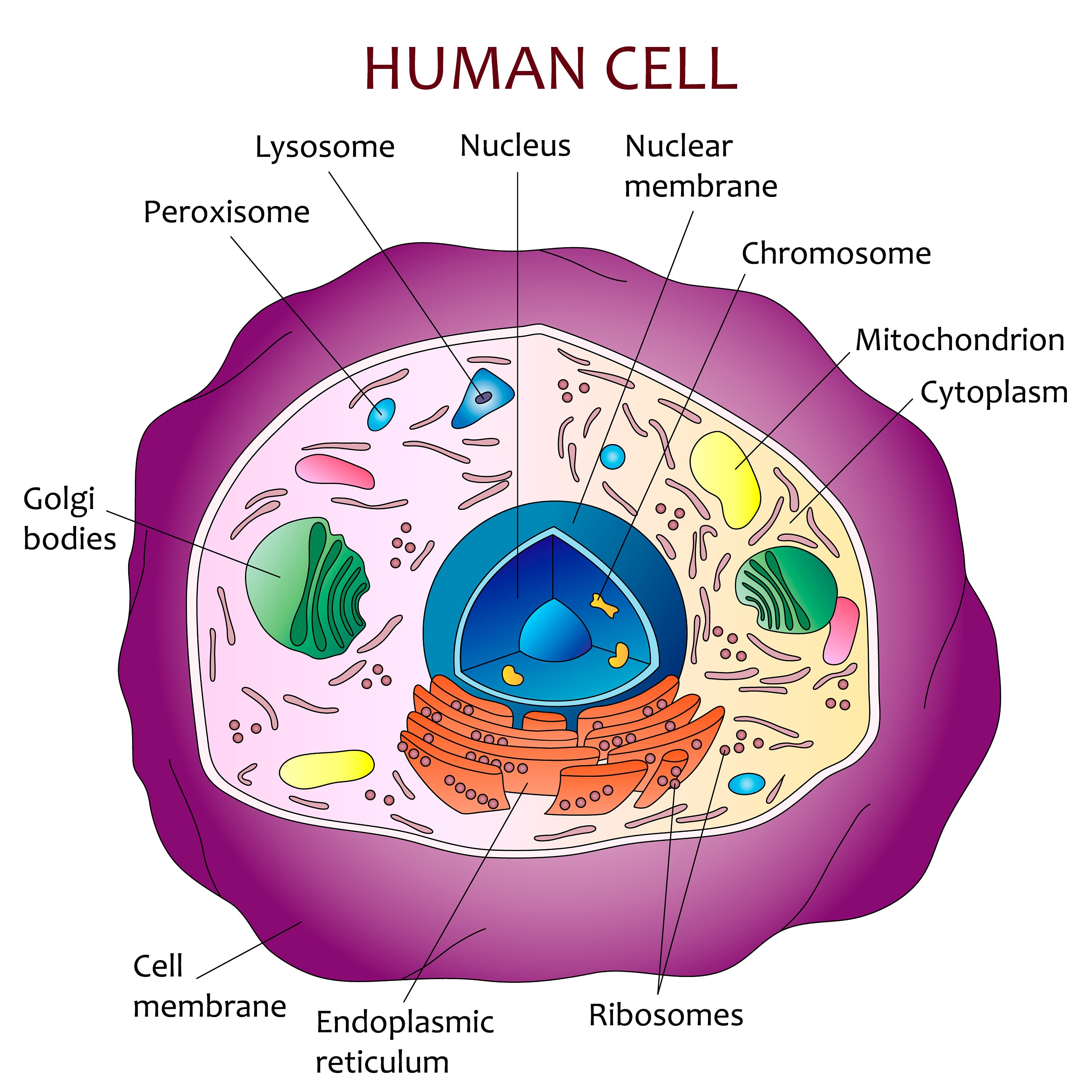
A brief explanation of the different parts of an animal cell along with a well-labelled diagram is mentioned below for reference. Also Read Different between Plant Cell and Animal Cell Well-Labelled Diagram of Animal Cell The Cell Organelles are membrane-bound, present within the cells.
Structure of cell Cell structure and functions, Class 8
A medium-sized circular cell part that has squiggly lines inside is labeled nucleus. The outermost part of the cell, which is shown as an outline of the cell, is labeled cell membrane. On the right is a four-sided figure with rounded corners that represents a plant cell. The cell contains many cell parts with different shapes.

Labeled Animal Cell Diagram
The nucleoid and some other frequently seen features of prokaryotes are shown in the diagram below of a cut-away of a rod-shaped bacterium. Image of a typical prokaryotic cell, with different portions of the cell labeled. _Image credit: modified from "Prokaryotic cells: Figure 1" by OpenStax College, Biology, CC BY 3.0_

South Pontotoc Biology Plant and Animal Cell Diagrams
Animal Cell Anatomy. The cell is the basic unit of life. All organisms are made up of cells (or in some cases, a single cell). Most cells are very small; in fact, most are invisible without using a microscope. Cells are covered by a cell membrane and come in many different shapes.

Plant Cell Diagram Labeled 3D / Plant Cell Stock Illustrations 8 546
Cell Diagrams with Labelling Activity. I've created two interactive diagrams for an upcoming open textbook for high-school level biology. The cell structure illustrations for these diagrams were generated in BioRender. Both diagrams feature a drag-and-drop labelling activity created with H5P here on Learnful.

Pin by james paterson on A (growing) list of people, places and things
Labeled diagram of a typical animal cell Nucleus. The nucleus contains all the genetic material in a cell. This genetic information is called deoxyribonucleic acid (DNA). DNA contains all the instructions for making proteins, which control all of the body's activities. Therefore, the nucleus is like the manager's office of the cell.
Cell Membrane Images Worksheet Answers
Plasma Membrane - Also known as the cell membrane, the plasma membrane is a selectively permeable wall that separates the cell interior from the outside environment. Ribosomes - The ribosomes are made of protein and RNA. They convert genetic material into protein.

Draw A Labelled Diagram Of Animal Cell Describe The Structure And
Figure 6.4 Animal cell mitosis is divided into five stages—prophase, prometaphase, metaphase, anaphase, and telophase—visualized here by light microscopy with fluorescence. Mitosis is usually accompanied by cytokinesis, shown here by a transmission electron microscope. (credit "diagrams": modification of work by Mariana Ruiz Villareal; credit "mitosis micrographs": modification of work by.
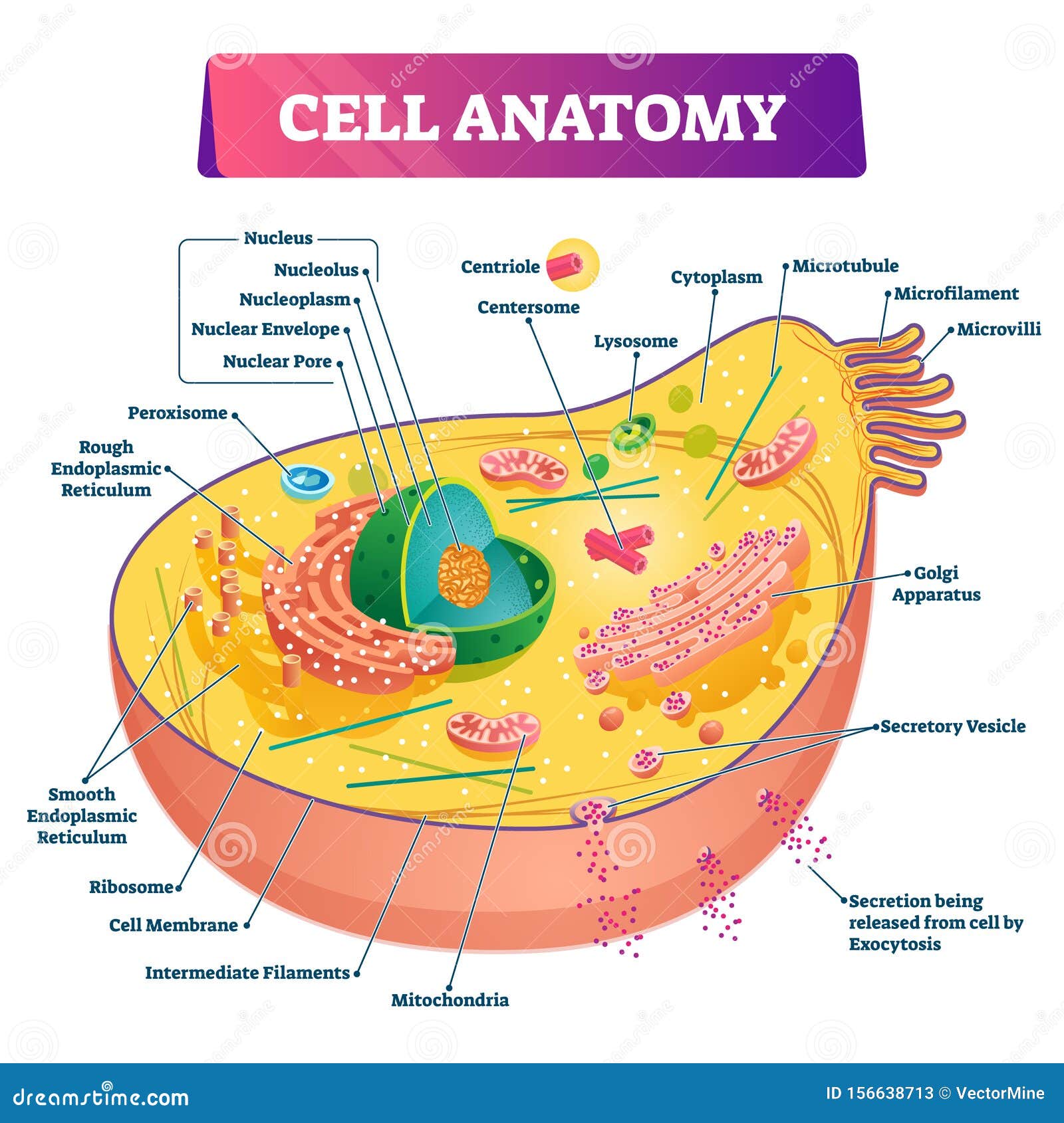
Cell Anatomy Vector Illustration. Labeled Educational Structure Diagram
They're one of two major classifications of cells - eukaryotic and prokaryotic. They're also the more complex of the two. Eukaryotic cells include animal cells - including human cells - plant cells, fungal cells and algae. Eukaryotic cells are characterized by a membrane-bound nucleus.
Education 645 High School Biology
Cell diagram labeled Cell diagram unlabeled Learn faster with interactive cell quizzes Sources + Show all What are the parts of a cell? There exist two general classes of cells: Prokaryotic cells: Simple, self-sustaining cells (bacteria and archaea) Eukaryotic cells: Complex, non self-sustaining cells (found in animals, plants, algae and fungi)

View 20 All Parts Of An Animal Cell Labeled Eporali Wallpaper
What exactly is its job? The plasma membrane not only defines the borders of the cell, but also allows the cell to interact with its environment in a controlled way. Cells must be able to exclude, take in, and excrete various substances, all in specific amounts.
Plant Cell Diagram Labeled EdrawMax Template
cell, in biology, the basic membrane-bound unit that contains the fundamental molecules of life and of which all living things are composed.A single cell is often a complete organism in itself, such as a bacterium or yeast.Other cells acquire specialized functions as they mature. These cells cooperate with other specialized cells and become the building blocks of large multicellular organisms.
Human cell diagram Etsy
Animal cells are eukaryotic cells, meaning they possess a nucleus and other membrane-bound organelles. Unlike plant cells, animal cells do not have cell walls, allowing for more flexibility in shape and movement. A plasma membrane encloses the cell contents of both plant and animal cells, but it is the outer coating of an animal cell.
